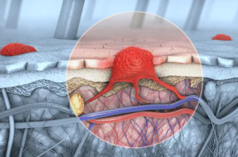
 In the deepest part of the skin, just above the dermis, there are cells called melanocytes. Melanocytes produce skin pigment or color. Melanoma begins when healthy melanocytes change and grow uncontrollably to form cancerous tumors. Melanoma is a tumor derived from the malignant transformation of melanocytes. Melanoma has a high degree of malignancy, it mostly occurs in the skin and it can also occur in different parts or tissues such as the mucous membrane, the uvea, and the pia mater. Melanoma has a high degree of malignancy, but its morbidity and mortality are relatively low. Some factors may increase the risk of melanoma, such as excessive UV exposure, family history of melanoma, etc.
Diagnosis of Melanoma
The diagnosis methods of melanoma are as follows:
- Physical examination. A preliminary diagnosis can be made by focusing on the skin, local and regional lymph node examination.
- Histopathological examination. This is a necessary step in the diagnosis of melanoma. The biopsy of the lesion is histologically confirmed. For patients with no distant metastasis in the initial clinical diagnosis, the melanoma is usually completely removed by the biopsy.
- Imaging diagnosis. For diagnosis and staging, the basic strategies include regional lymph node ultrasound, chest X-ray, CT, pelvic ultrasound, enhanced CT, MRI, whole-body bone scan, enhanced MRI, and sometimes whole-body PET-CT.
- Laboratory examination. Blood routine, liver and kidney function and lactate dehydrogenase (LDH), these indicators are mainly to prepare for subsequent treatment and to understand the prognosis.
- Immunohistochemical testing. Detect the characteristic markers of melanocytes (S-100 protein, SOX-10, Melan A, HMB45, tyrosinase, etc.) to identify various tumors such as melanoma, sarcoma and lymphoma. Immunohistochemical markers including KI-67 and Cyclin D1 were detected to assist the differential diagnosis of skin melanoma and benign melanocyte nevus.
- DNA Testing. There are many genetic changes in the occurrence and development of melanoma. Genetic testing is helpful in the diagnosis of some difficult cases, and can also predict the efficacy of targeted therapy drugs and guide clinical treatment. It is recommended that all melanoma patients undergo genetic testing for BRAF, c-Kit and NRAS before treatment.
Advantages of Our Products
Creative Biogene's products are mainly for the diagnosis of melanoma by genetic testing, including a series of mutation test kits, which are based on the polymerase chain reaction (PCR) detection method and use allele-specific primers to identify the presence of BRAF, NRAS, and c-Kit mutations in multiple reactions.
Creative Biogene’s skilled scientists have experience in developing outstanding products in the field of melanoma diagnosis. We will uphold the belief of pursuing high quality and the spirit of professionalism to provide you with our optimal products. You can choose us without hesitation.
Please contact us for more details.
Reference
- Sperduto, P W, et al. (2017). "The prognostic value of BRAF, C-KIT, and NRAS mutations in melanoma patients with brain metastases." International Journal of Radiation Oncology Biology Physics. 98.5: 1069-1077.
For research use only. Not for any other purpose.
 USA:
USA:  Europe:
  Email:
  Consultation


 Diagnostic Development


