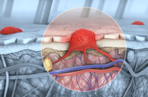
 In the deepest part of the skin, just above the dermis, there are cells called melanocytes. Melanocytes produce skin pigment or color. Melanoma begins when healthy melanocytes change and grow uncontrollably to form cancerous tumors. Melanoma is a tumor derived from the malignant transformation of melanocytes. Melanoma has a high degree of malignancy, it mostly occurs in the skin and it can also occur in different parts or tissues such as the mucous membrane, the uvea, and the pia mater. Melanoma has a high degree of malignancy, but its morbidity and mortality are relatively low. Some factors may increase the risk of melanoma, such as excessive UV exposure, family history of melanoma, etc.
The diagnosis methods of melanoma are as follows:
Creative Biogene's products are mainly for the diagnosis of melanoma by genetic testing, including a series of mutation test kits, which are based on the polymerase chain reaction (PCR) detection method and use allele-specific primers to identify the presence of BRAF, NRAS, and c-Kit mutations in multiple reactions.
Creative Biogene’s skilled scientists have experience in developing outstanding products in the field of melanoma diagnosis. We will uphold the belief of pursuing high quality and the spirit of professionalism to provide you with our optimal products. You can choose us without hesitation.
Please contact us for more details.
Reference
| Cat# | Product Name | Product Type | Inquiry |
|---|---|---|---|
| C0540A | Pinin polyclonal antibody | Antibody | Inquiry |
| C0541A | PRAME polyclonal antibody | Antibody | Inquiry |
| C0542A | RAB38 polyclonal antibody | Antibody | Inquiry |
| C0543A | SILV polyclonal antibody | Antibody | Inquiry |
| C0544A | SLC45A2 polyclonal antibody | Antibody | Inquiry |
| C0545A | Syntenin 2 polyclonal antibody | Antibody | Inquiry |
| C0546A | Syntenin-1 polyclonal antibody | Antibody | Inquiry |
| C0547A | ZNF654 polyclonal antibody | Antibody | Inquiry |
Copyright © 2026 Creative Biogene. All rights reserved.